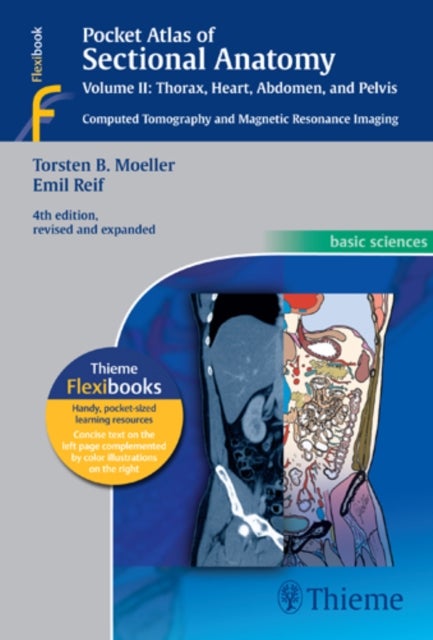
Pocket Atlas of Sectional Anatomy, Vol. II: Thorax, Heart, Abdomen and Pelvis av Torsten Bert Moeller, Emil Reif
589,-
<p>This comprehensive, easy-to-consult pocket atlas is renowned for its superb illustrations and ability to depict sectional anatomy in every plane. Together with its two companion volumes, it provides a highly specialized navigational tool for all clinicians who need to master radiologic anatomy and accurately interpret CT and MR images.</p><p>Special features of <em>Pocket Atlas of Sectional Anatomy</em>:<ul><li>Didactic organization in two-page units, with high-quality radiographs on one side and brilliant, full-color diagrams on the other<li> Hundreds of high-resolution CT and MR images made with the latest generation of scanners (e.g., 3T MRI, 64-slice CT)<li> Color-coded schematic drawings that indicate the level of each section <li> Consistent color coding, making it easy to identify similar structures across several slices</li></ul><p>Updates for the 4th edition of Volume II:</p><ul><li>CT imaging of the chest and abdomen in all 3 planes: axial, sagittal, and coronal<li> New ba

Relaterte produkter
Vis flereVi har valgt ut en rekke interessante produkter i samme kategori som Pocket Atlas of Sectional Anatomy, Vol. II: Thorax, Heart, Abdomen and Pelvis av Torsten Bert Moeller, Emil Reif. Hvis du ikke finner noe interessant her kan du enkelt klikke på “vis flere”.


Produktinformasjon
- Alle prisene nevnt ovenfor er oppgitt i Norske kroner.
- Dette produktet er tilgjengelig hos Norli NO.
- Hos Norli NO kan du kjøpe Pocket Atlas of Sectional Anatomy, Vol. II: Thorax, Heart, Abdomen and Pelvis av Torsten Bert Moeller, Emil Reif for kun 589,-.
- Den laveste prisen på Pocket Atlas of Sectional Anatomy, Vol. II: Thorax, Heart, Abdomen and Pelvis av Torsten Bert Moeller, Emil Reif ble registrert 17. februar 2025 kl. 14:48.
Prisutvikling
Er den nåværende prisen et godt tilbud?
Grafen over prisutviklingen viser den laveste prisen over tid, eksklusiv fraktkostnader.
Prishistorikk for Pocket Atlas of Sectional Anatomy, Vol. II: Thorax, Heart, Abdomen and Pelvis av Torsten Bert Moeller, Emil Reif
Laveste pris
589,-
17 feb. 2025
Høyeste pris
589,-
17 feb. 2025