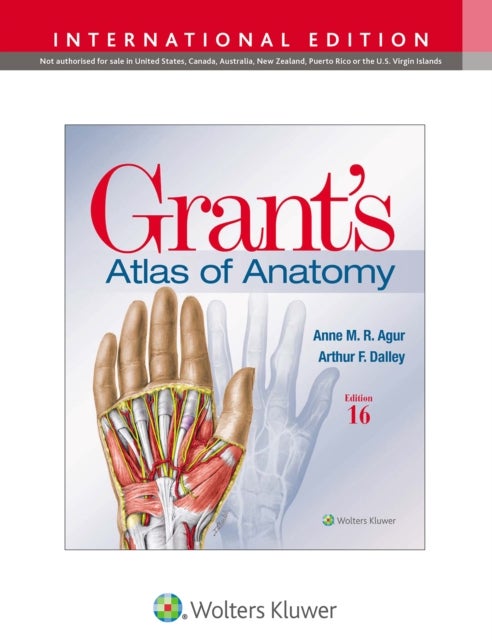
Grant's Atlas of Anatomy av Anne M. R. B.Sc. (OT) M.Sc PhD Agur, Arthur F. PhD FAAA Dalley II
869,-
Illustrations drawn from real specimens, presented in surface-to-deep dissection sequence, set <I>Grant’s Atlas of Anatomy</I> apart as the most accurate illustrated reference available for learning human anatomy and referencing in dissection lab. A recent edition featured re-colorization of the original Grant’s Atlas images from high-resolution scans, also adding a new level of organ luminosity and tissue transparency. The dissection illustrations are supported by descriptive text legends with clinical insights, summary tables, orientation and schematic drawings, and medical imaging. <ul><li> Renowned, high-resolution, dynamically colored illustrations organized in dissection sequence enable the formation of 3D constructs for each body region and provide detailed, realistic reference during dissection. </li><li> Tables detail muscles, vessels, and other anatomic information in an easy-to-use format ideal for review and study. </li><li> Enhanced medical imaging includes mor

Relaterte produkter
Vis flereVi har valgt ut en rekke interessante produkter i samme kategori som Grant's Atlas of Anatomy av Anne M. R. B.Sc. (OT) M.Sc PhD Agur, Arthur F. PhD FAAA Dalley II. Hvis du ikke finner noe interessant her kan du enkelt klikke på “vis flere”.


Produktinformasjon
- Alle prisene nevnt ovenfor er oppgitt i Norske kroner.
- Dette produktet er tilgjengelig hos Norli NO.
- Hos Norli NO kan du kjøpe Grant's Atlas of Anatomy av Anne M. R. B.Sc. (OT) M.Sc PhD Agur, Arthur F. PhD FAAA Dalley II for kun 869,-.
- Den laveste prisen på Grant's Atlas of Anatomy av Anne M. R. B.Sc. (OT) M.Sc PhD Agur, Arthur F. PhD FAAA Dalley II ble registrert 17. februar 2025 kl. 14:48.
Prisutvikling
Er den nåværende prisen et godt tilbud?
Grafen over prisutviklingen viser den laveste prisen over tid, eksklusiv fraktkostnader.
Prishistorikk for Grant's Atlas of Anatomy av Anne M. R. B.Sc. (OT) M.Sc PhD Agur, Arthur F. PhD FAAA Dalley II
Laveste pris
869,-
17 feb. 2025
Høyeste pris
869,-
17 feb. 2025